The Stroke Advocacy Network (SAN) is the voice of the stroke community.
SAN members impact public policy outcomes by influencing decision makers through writing letters, signing petitions, meeting with their legislators, and much more.
Become an advocate, Go Here
SuperTracker
Find out what and how much to eat.
Personalize your experience by creating your profile
Get a plan tailored for you.
Track and score diet and physical activity and receive tips for healthy changes.
Get your personalized nutrition and physical activity plan.
Track your foods and physical activities to see how they stack up.
Get tips and support to help you make healthier choices and plan ahead.
Start Your Profile
Survivors of heart disease and stroke are not alone. In fact, many of them are sharing their stories to reach out to the community. They share their stories to champion hope and support to help us fight the devastation of heart disease and stroke.
24 million Americans have COPD. Are you one of the missing millions?
It takes less than a minute to find out if you are at risk for COPD.
Take The Screener Click Here
This web site maintains a huge list of medical statistics including misdiagnosis and prevalence/incidence data for hundreds of medical conditions.
Learn More
The American Red Cross prevents and alleviates human suffering in the face of emergencies by mobilizing the power of volunteers and the generosity of donors. Donate and/or Join
VAERS provides a nationwide mechanism by which adverse events following immunization may be reported, analyzed, and made available to the public. Go Here
About 3 million adults in the US are infected with the hepatitis C virus, most are baby boomers.
Baby boomers, anyone born from 1945 through 1965, should get tested for hepatitis C. Read More
STD - diseases and related conditions
Prevention and Treatment
How You Can Prevent STDs
Basic ways to prevent all STDs. Learn More
Battles are public health priorities with large-scale impact on health and known effective strategies to address them. Read More
Get involved in the fight against stroke
Youre the Cure is the AHA/ASA network of people who advocate for policies
that support a heart disease and stroke-free America. Learn More
This tool provides instant access to state level health and demographic data about adults with disabilities. state profiles,Maps, data tables, state profiles, and much more. Go Here
Methicillin-resistant Staphylococcus aureus (MRSA) is a bacteria that is resistant to many antibiotics. More Info
A guide to help you quit smoking, including reasons to quit, steps to quit, tips on handling cravings, medications that can help, and what to do if you slip. Try It
The Stroke Help Line is a volunteer-staffed call center for survivors, caregivers, family members and those who have experienced stroke in their lives.
This service can be accessed through 1-800-STROKES (787-6537), menu option 3. Visit Site
Lung Cancer Screening Tool.
Lung Cancer Screening Saves Lives. No One Deserves Lung Cancer. Today; lung cancer is the #1 cancer killer in America.
Should you be screened for lung cancer? Find Out
Nationally Accepted Online CPR Training And Certification
Training courses designed by a team of U.S board certified and licensed medical doctors.
Designed to help the general public as well as teachers, nursing home workers, daycare workers, and other medical workers gain the CPR skills and knowledge needed to help save a life. Check Us Out
No doubt about it the heart plays about the most important role not only in the functions of the body and the things you do but for the health and well being of your entire body.
The heart needs and deserves your utmost attention daily for your well being. The health and condition of your heart depends on you.
Here I will try to discuss and/or provide links to information about the many functions of the heart and the many diseases and conditions that affect the heart and/or the result of a malfunction of the heart, especially the Heart Attack and some of the causes for heart problems and the things you can do to maintain a healthy heart.
The Heart Attack, Heart Surgery and Heart Failure will be covered in separate sections of this site. You may navigate to them from one of the menus at the top left side or bottom of each page.
You can also find information about the Heart at the Medical Links Page and the Frequently Asked Questions and Answers Page
Here are fifteen (15) PDF text articles about The Heart, click and "Select" one and click "Go".
You can open and read your selection or download and save it to your computer to read and/or reference later.
Open & View and/or Save & Download
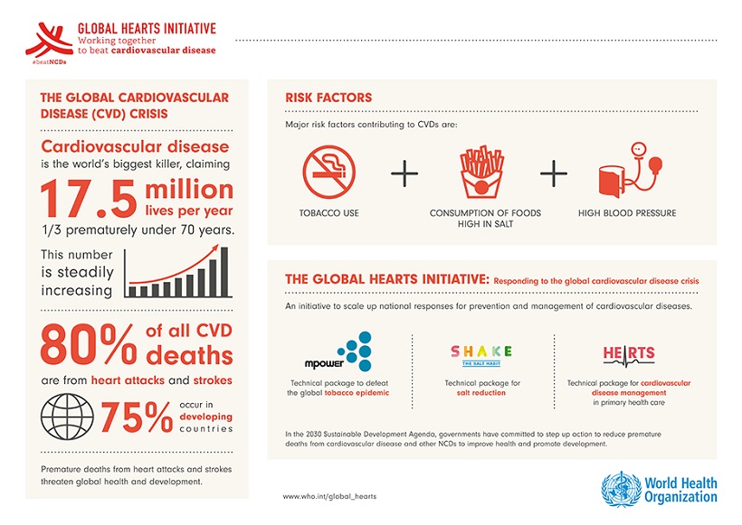
Cardiovascular Disease By Country
Cardiovascular diseases, commonly referred to as heart disease or stroke, are the number 1 cause of death globally. The factsheets below provide a snapshot from different corners of the world, demonstrating the scale of the problem and what governments are doing to respond.
The Heart
Throughout human history the rhythmic beat of the heart has quintessentially represented life. Until the advent of the heartlung machine, the lack of a heart beat, unless reversed within a few minutes, invariably signalled death. The beat of our own heart can be apparent to us in the pulse felt, or seen, at various parts of the body, occasionally heard or because of an unusual rhythm or skipped beat noticeable in the chest.
The heart is a hollow muscular organ. It acts as the prime mover for the circulation of the blood and the maintenance of the blood pressure. A certain volume of blood is delivered with each beat, and a further key aspect is the pressure at which this flow is delivered. Vital functions such as those of lungs and kidneys, or the exchange of components of the blood and tissue fluid at the capillaries, are critically dependent on the pressure achieved within the circulatory system.
Anatomy
The heart comprises a series of blood-filled chambers; the walls are composed virtually entirely of muscle cells of a type unique to the heart (cardiac myocytes). The heart is actually two double pumps acting in series; there are four chambers in all. The right side receives blood returning from the entire body (in the great veins) and pumps it into the pulmonary artery, which supplies only the alveoli (gas exchange sites) in the lungs. The left side receives blood from the lungs and pumps it into the aorta, the largest artery. (The heart is generally illustrated as seen from the front, so left and right appear mirrored).
The aorta branches to form the arterial tree that supplies blood to the whole body. The heart, appropriately, is itself the first organ supplied with blood from the aorta. The coronary arteries open from the beginning of the aorta and take blood to all parts of the heart tissue. Each side of the heart has an upper chamber, the atrium (plural: atria), into which the veins drain. They serve as antechambers to the respective ventricles, the thicker-walled chambers that lie below them.
Atria and Valves
The arrangement of one-way valves and the prevailing pressures mainly determine blood flow from veintoatriumto-ventricle during the cyclic activity of the heart beat, but some pumping of blood by the atria into the ventricles also occurs. The valves preventing back-flow from ventricle to atrium are tough, parachute-like structures partly anchored in the connective tissue plate which forms the physical union of the ventricular and atrial portions of the heart. Their free edges are restrained by several papillary muscles. These are slim extensions from the inner wall of the ventricles, each with a tendinous end fused with the valve; acting like parachute cords, they prevent the valve being pushed through into the atrium as its flaps become filled when the ventricle contracts and puts pressure on its contents. The mitral (or bicuspid) valve on the left side has two flaps, and the tricuspid valve on the right has three. The parachutes press together forming a complete closure preventing regress of blood into the respective atrium whence it came. Instead, when the pressure has risen sufficiently, blood is directed into the pulmonary artery and the aorta through one-way valves which separate them from, and prevent back-flow into, their respective ventricles.
The Heart Beat
The heart beats between 60 and 220 times per minute in a typical young adult; 40 to 50 million beats per year. The rate alters, often rather obviously, according to one's state of physical and mental activity. This results in pumping over 3 million litres of blood (per year) through the body and an equal volume through the lungs. The pump work done by the heart is equivalent to lifting a 1 kg weight to about twice the height of Mount Everest each day. This level of persistent, rhythmic, and decidedly dynamic activity may provoke a sense of awe, although it is hardly more remarkable than the prosaic activity of every other organ except in its absolute necessity to keep at it! We will first consider the electrical processes of the heart since, like many muscles, it is triggered into activity (contraction, the heart beat) by an electrical wave. This section is followed by consideration of contraction itself.
Electrical Aspects
The left and right atria beat virtually simultaneously and then, after a fraction of a second's delay, both ventricles contract. Electrical activity, as in most other muscles, triggers the contraction. This activity arises not from excitatory nerve fibres, but spontaneously within the heart itself from a small clump of pacemaker cells near the point where the vena cava joins the right atrium: the sino-atrial (SA) node. The electrical wave, or action potential, spreads across the heart from cell to cell. This spread is made possible because each heart cell is connected to its immediate neighbours at several contact regions which offer a relatively low resistance to the flow of electrical current. All the muscle cells of the heart are thus electrically linked together. This means that the activity spreads as a wave, its direction determined by the cell-to-cell couplings available. It also means that, as far as we know, every cardiac myocyte is active at some stage during every heart beat. The muscle cells of the atria and ventricles only make electrical contact in one small region, the atrio-ventricular (AV) node at the centre of the heart. Thus, activity follows a predictable, regular path, across the right and left atria, through the AV node, along specialized faster-conducting heart cells (Purkinje fibres) on the internal face of the muscular wall between the two ventricles (interventricular septum), and thence through the substance of both ventricles. Heart cells, like other electrically excitable cells, become inexcitable (refractory) for a brief period after each action potential. Consequently, once the wave has passed right through the ventricles it ceases, since there are no non-refractory cells available to excite. A new wave is spontaneously initiated at the pacemaker region.
Contractile (mechanical) Aspects
All the heart muscle cells are thus electrically excited and it is this that triggers them to contract. The wave of contraction, therefore, follows the same sequence: atria first, then ventricles. The electrical activity triggers an abrupt rise in the concentration of free calcium ions inside the cells a common feature in signalling contraction in muscle of every type. The calcium ions required are derived in part by influx from the extracellular fluid, in part by release from intracellular stores in the sarcoplasmic reticulum. The influx is through calcium-selective channels in the surface membrane which are opened by the depolarization. The influx itself transiently promotes further influx, and also triggers the release of more calcium from the intracellular store.
In each ventricle, as the muscular walls contract (develop tension and shorten) they press upon the blood they enclose. The pressure rises and the AV valve fills out and closes. At this stage of the cycle, the exit valve into the relevant artery (pulmonary artery or aorta) is also closed because the pressure in the arteries is higher than that in the ventricles. Temporarily, each ventricle is thus a closed chamber, it can neither lose nor gain blood, so pressure rises quickly until it exceeds that in the exit artery; the exit valve is then pushed open and blood is ejected, squirted from the ventricles as their muscular walls continue to shorten. The pressure at which the valve opens is much higher on the left side than on the right side, in accordance with the higher blood pressure in the aorta and its branches than in the pulmonary artery and its branches. The resistance offered by the lungs to blood flow is much less than that by the body generally; thus the pressures required of the right ventricle can be lower, yet achieve the same flow rate. Both ventricles eject the same volume of blood (the stroke volume): in the adult heart, about 70 ml (half a teacup) which is half or less of the volume it contained. As action potential finishes, the intracellular calcium concentration has already started to reduce again: some calcium is being pumped back into the store, and some is leaving the cell by an ion exchange process. With the raised calcium concentration signal thereby removed, the force of contraction quickly wanes in the muscle, so ventricular pressure falls. The elasticity of the arteries, which were dilated when blood was ejected into them, now ensures that a higher pressure is sustained in them than in the rapidly relaxing ventricles (the garden hose effect, familiar to those who have turned off a hose-pipe supply tap only to see water continue squirting as the elastic pipe collapses). The respective exit valves are thus pushed closed again, preventing reflux into the ventricles. Blood pressure, therefore, falls more slowly in the arteries than in the ventricles. At this stage about 90 ml of blood remains in each ventricle. Pressure continues to fall quickly until it is below that in the atria. Thus, the AV valves are pushed open, allowing blood to flow from the atria into the ventricles topping them up with more blood. (Despite the appearance in some published schematic diagrams and cartoon sequences, at all stages of the heart beat the chambers are full of blood. It is the enclosed volume which changes, depending on the tension and elasticity of the muscular walls and the status of the inlet and outlet valves.)
The return of the ventricle to its resting shape between beats is due to its own elasticity. Like a squeezed sponge or hollow rubber ball, this significantly sucks blood from the atria, thereby contributing to its own filling. The reduction of this factor in old age or its enhancement by athletic training have a major effect on overall cardiac function. These effects are analogous to problems associated with stiff inelastic valves which perhaps more obviously compromise effective flow in and out of the chambers of the heart.
The state when the heart is contracting is termed systole (sis'-toe-lee); the relaxed state is termed diastole (di-a'-stoe-lee).
Control of Pump Function
The cardiac output is the volume of blood pumped per minute by each ventricle some 5 litres/minute at rest and is simply the product of heart rate and stroke volume. Cardiac output will thus alter if either varies. The stroke volume is in turn influenced by cardiac filling and by the contractility of the cardiac muscle itself its intrinsic ability to contract (shorten and/or produce tension).
Heart Rate
The earliest human hunters will have noticed, like later horror film makers, that even when removed from the body, the heart continues to beat for a time. Other organs also continue to live, but their activity is hardly as impressive as that of the heart.
Because all the cells of the heart are electrically connected to their neighbours, the whole behaves as a unit. Most regions are inactive, unless artificially stimulated. The activity of the regions with the property of firing spontaneously is conducted to all their inactive neighbours, so they act as pacemakers. The inherent pacemaker firing rate, typically about 100 per minute, is influenced by nerve actions of the autonomic nervous system: sympathetic nerves release noradrenaline which increases rate, and parasympathetic (vagus) nerve fibres release acetylcholine which slows the rate. Heart rate typically varies between 60 per minute (in deep sleep) to approaching 200 per minute (during brief bursts of maximal exercise). The normal resting rate while sitting, relaxed, is about 70 per minute, but shows wide variation amongst entirely healthy individuals. (In one university class of 350 twenty-year-old students, the range was 48 to 90 per minute.) One common feature is a marked variation within the breathing cycle: breathing in usually increases the rate. Physical fitness, particularly that associated with endurance rather than muscle strength, is often associated with a low resting rate. Extremes such as the tennis player Bjorn Borg, or the professional cyclist Miguel Indurain, with resting values in the low 30s per minute, are well known. Young children have higher resting rates; whilst still in the womb, a baby will have a rate of 120 to 160 beats per minute; it is often reported that rates above 140 indicates a female baby, but there are more reliable tests!
Cardiac Filling
Filling reflects the flow of blood back into the heart (venous return from the lungs and the body). William Harvey observed that the presence of values requires that blood in the larger veins can only flow towards the heart, the key to recognizing that blood circulates. Amongst other factors, the extent of muscular activity, breathing movements, and body positions (standing, lying, arms or legs raised) all affect the rate of return of blood to the heart. Cardiac muscle shows the unusual property that, within limits, it contracts more powerfully when starting from stretched lengths, so that the ventricle empties more forcibly when it is filled more than usual. This is achieved at trivial extra metabolic cost; the efficiency of pumping thus increases as output increases; surely a paradigm for productivity gains. This property allows the heart to compensate automatically when the volume of blood within it at the start of the beat (the end diastolic volume) is greater than previously, by pumping more forcefully, thus ejecting a larger volume. This feature is termed Starling's Law of the Heart, after one of its discoverers.
Contractility
It is obvious that an intrinsically stronger heart will be able to eject blood more forcefully and more completely. Unlike our voluntary (skeletal) muscles, the strength of heart muscle can vary quickly, even from one beat to the next. This is because it is sensitive to chemical influences (especially of adrenaline/noradrenaline) and electrical influences that can rapidly modify the intracellular processes that underlie contraction. Additionally, as with voluntary muscle, the extent of growth and development of the heart muscle will affect the overall strength of the organ; athletes generally have thicker heart walls which match the larger muscles in their thicker limbs. A normal, sudden increase in contractility is associated with the onset of physical activity or even with its anticipation; this is signalled to the heart, along with the increase in heart rate, by activity in the sympathetic nerve fibres which release noradrenaline. The combination of higher rate and stronger, more rapid contraction tends to match cardiac output to the increased demands for blood flow to the exercising muscles.
The Heart of the Matter and the Matter of the Heart
The control systems which influence the heart rate and strength of beating are the same as those implicated in such apparently diverse processes as blushing, breathing rate, sexual arousal, mental stress, or alertness. These links seem to have been recognized by our forebears in advance of the definitive precision of the discoveries of cardiovascular physiology. Poets report that hearts leap, hearts are strong, hearts are united, hearts are hot, heart strings are plucked, hearts are in the mouth, hearts become feeble, hearts are chilled, hearts tremble, and hearts are broken. In human history, the nature of the circulation of the blood and the (quite literally) central role of the heart in this system are still recent discoveries, even though they rank with the very earliest of the truly modern scientific method. Nevertheless, the heart (with perhaps the eye) is the organ most quoted in literature and song to define the essential qualities of life and even its very presence.
The ready perception of the action of the heart, its racing rate when we are excited or surprised, aroused or shocked, the shallow, rapid beat encountered in feverish poor health, the occasional irregularity of beat that can concern us all (often, thankfully, quite unnecessarily), together form the shared heart experiences of mankind that writers and poets have ever drawn upon. We are generally blissfully unaware of the other hives of metabolic industry that contribute to our physiology. The liver, the thyroid, the hypothalamus, the pituitary, the spleen, the pancreas, not one of these is dignified with a property recognizable to their owners. It is surely the literal vitality of the heart's rhythmic beating, the recognition of its link to the movements of blood, the necessary identity between this continual activity and life itself (outside an operating theatre) that validates the continuing truth of poetic notions of "The Heart".
Source: Encyclopedia.com
Source: WebMD
13 Surprising Things That Hurt Your Heart
Begin >>
All About Heart Rate (Pulse) and Target Heart Rates
What should you know about your heart rate:
Even if youre not an athlete, knowledge about your heart rate can help you monitor your fitness level and it might even help you spot developing health problems.
Your heart rate, or pulse, is the number of times your heart beats per minute. Normal heart rate varies from person to person. Knowing yours can be an important heart-health gauge.
As you age, changes in the rate and regularity of your pulse can change and may signify a Heart Condition or other condition that needs to be addressed.
Where is it and what is a normal heart rate?
The best places to find your pulse are the:
wrists
inside of your elbow
side of your neck
top of the foot
To get the most accurate reading, put your finger over your pulse and count the number of beats in 60 seconds.
Your resting heart rate is the heart pumping the lowest amount of blood you need because youre not exercising. If youre sitting or lying and youre calm, relaxed and arent ill, your heart rate is normally between 60 (beats per minute) and 100 (beats per minute).
But a h eart rate lower than 60 doesnt necessarily signal a medical problem. It could be the result of taking a drug such as a BetaBlocker. A lower heart rate is also common for people who get a lot of physical activity or are very athletic. Active people often have lower heart rates because their heart muscle is in better condition and doesnt need to work as hard to maintain a steady beat.
Moderate physical activity doesnt usually change the resting pulse much. If youre very fit, it could change to 40. A less active person might have a heart rate between 60 and 100. Thats because the heart muscle has to work harder to maintain bodily functions, making it higher.
How Other Factors Affect Heart Rate
Air temperature: When temperatures (and the humidity) soar, the heart pumps a little more blood, so your pulse rate may increase, but usually no more than five to 10 beats a minute.
Body position: Resting, sitting or standing, your pulse is usually the same. Sometimes as you stand for the first 15 to 20 seconds, your pulse may go up a little bit, but after a couple of minutes it should settle down. Emotions: If youre stressed, anxious or extraordinarily happy or sad your emotions can raise your pulse.
Body size: Body size usually doesnt change pulse. If youre very obese, you might see a higher resting pulse than normal, but usually not more than 100.
Medication use: Meds that block your adrenaline (beta blockers) tend to slow your pulse, while too much thyroid medication or too high of a dosage will raise it.
When To Call Your Doctor
If youre on a beta blocker to decrease your heart rate (and Lower Blood Pressure) or to control an abnormal rhythm (Arrhythmia), your doctor may ask you to monitor and log your heart rate. Keeping tabs on your heart rate can help your doctor determine whether to change the dosage or switch to a different medication.
If your pulse is very low or if you have frequent episodes of unexplained fast heart rates, especially if they cause you to feel weak or dizzy or faint, tell your doctor, who can decide if its an emergency. Your pulse is one tool to help get a picture of your health.
Arrhythmias (Abnormal Heart Rhythms)
Target Heart Rates How do you get your heart rate on target?
When you work out, are you doing too much or not enough? Theres a simple way to know: Your target heart rate helps you hit the bulls eye. We dont want people to over-exercise, and the other extreme is not getting enough exercise, says Gerald Fletcher, M.D., a cardiologist and professor in the Mayo Clinic College of Medicine in Jacksonville, Fla.
First Things First
Before you learn how to calculate and monitor your target training heart rate, you have to know your resting heart rate. Your resting heart rate is the number of times your heart beats per minute while its at rest. You can check it in the morning after youve had a good nights sleep and before you get out of bed.
According to the National Institute of Health, the average resting heart rate:
for children 10 years and older, and adults (including seniors) is 60 - 100 beats per minute
for well-trained athletes is 40 - 60 beats per minute.
Hittin the Target
Now youre ready to determine your target training heart rate. As you exercise, periodically:
Take your pulse on the inside of your wrist, on the thumb side.
Use the tips of your first two fingers (not your thumb) to press lightly over the blood vessels on your wrist.
Count your pulse for 10 seconds and multiply by 6 to find your beats per minute. You want to stay between 50 percent to 85 percent of your maximum heart rate. This range is your target heart rate.
Know Your Numbers
This table shows estimated target heart rates for different ages. Your maximum heart rate is about 220 minus your age.
In the age category closest to yours, read across to find your target heart rate. Heart rate during moderately intense activities is about 50-69% of your maximum heart rate, whereas heart rate during hard physical activity is about 70% to less than 90% of the maximum heart rate.
The figures are averages, so use them as general guidelines.
Age
Target HR Zone 50-85%
Average Maximum HR, 100%
20 years
100-170 beats per minute
200 beats per minute
30 years
95-162 beats per minute
190 beats per minute
40 years
90-153 beats per minute
180 beats per minute
45 years
88-149 beats per minute
175 beats per minute
50 years
85-145 beats per minute
170 beats per minute
55 years
83-140 beats per minute
165 beats per minute
60 years
80-136 beats per minute
160 beats per minute
65 years
78-132 beats per minute
155 beats per minute
70 years
75-128 beats per minute
150 beats per minute
Important Note: A few high blood pressure medications lower the maximum heart rate and thus the target zone rate. If you're taking such medicine, call your physician to find out if you need to use a lower target heart rate.
So whats in a number?
If your heart rate is too high, youre straining. So slow down. If its too low, and the intensity feels light or moderate/brisk, you may want to push yourself to exercise a little harder.
During the first few weeks of working out, aim for the lower ranger of your target zone (50 percent) and gradually build up to the higher range (85 percent). After six months or more, you may be able to exercise comfortably at up to 85 percent of your maximum heart rate.
Its not an absolute, but its a good tool to have, says Fletcher, who is also an American Heart Association volunteer. And if you dont know it, remember, if youre not able to carry on a conversation (while exercising), that may be a bit too much.
If you have a heart condition or youre in cardiac rehab, talk to a healthcare professional about what exercises you can engage in, what your target heart rate should be and whether you need to be monitored during physical activity. This will also help you to choose the types of physical activity that are appropriate for your current fitness level and health goals, because some activities are safer than others.
Miscellaneous strange, unusual, and seldom heard things about the Human Heart
36 Interesting Facts About The Human Heart
Source: FactRetriever.com
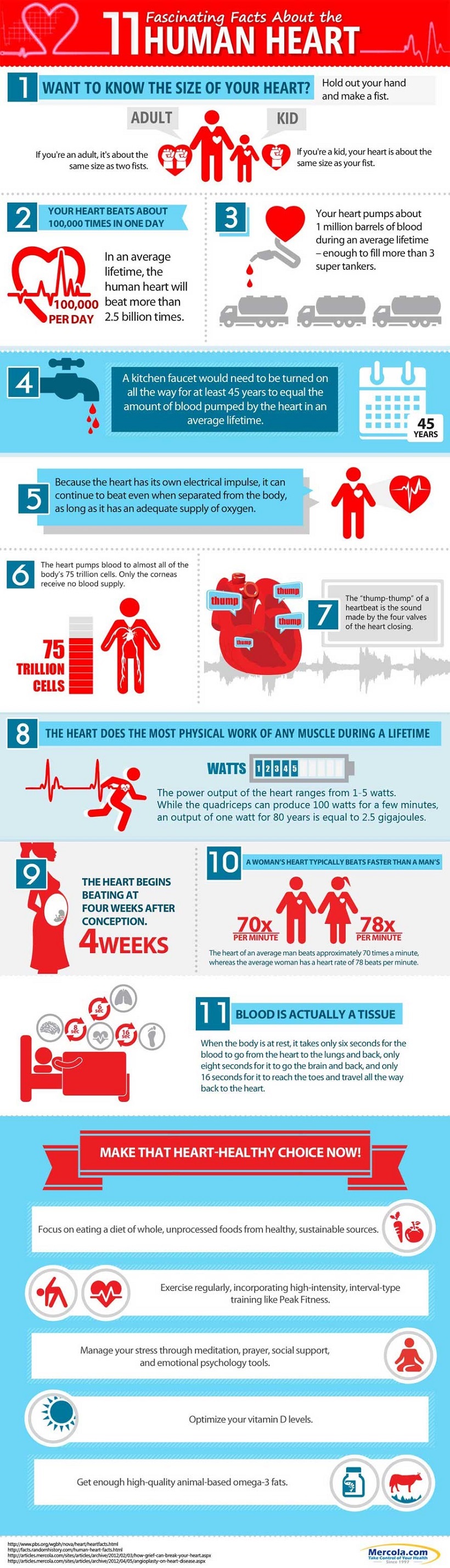
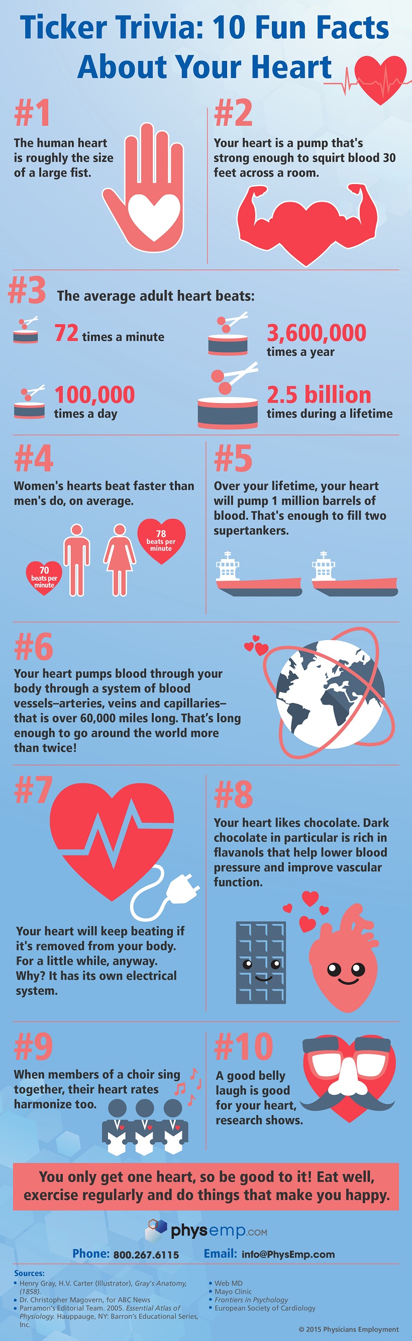
The average adult heart beats 72 times a minute; 100,000 times a day; 3,600,000 times a year; and 2.5 billion times during a lifetime.
Though weighing only 11 ounces on average, a healthy heart pumps 2,000 gallons of blood through 60,000 miles of blood vessels each day.
A kitchen faucet would need to be turned on all the way for at least 45 years to equal the amount of blood pumped by the heart in an average lifetime.
The volume of blood pumped by the heart can vary over a wide range, from five to 30 liters per minute.
Every day, the heart creates enough energy to drive a truck 20 miles. In a lifetime, that is equivalent to driving to the moon and back.
Because the heart has its own electrical impulse, it can continue to beat even when separated from the body, as long as it has an adequate supply of oxygen.
French physician Rene Laennec (1781-1826) invented the stethoscope when he felt it was inappropriate to place his ear on his large-buxomed female patients' chests.
The fetal heart rate is approximately twice as fast as an adults, at about 150 beats per minute. By the time a fetus is 12 weeks old, its heart pumps an amazing 60 pints of blood a day.
The heart pumps blood to almost all of the bodys 75 trillion cells. Only the corneas receive no blood supply.
During an average lifetime, the heart will pump nearly 1.5 million barrels of bloodenough to fill 200 train tank cars.
Five percent of blood supplies the heart, 15-20% goes to the brain and central nervous system, and 22% goes to the kidneys.
The thump-thump of a heartbeat is the sound made by the four valves of the heart closing.
The heart does the most physical work of any muscle during a lifetime. The power output of the heart ranges from 1-5 watts. While the quadriceps can produce 100 watts for a few minutes, an output of one watt for 80 years is equal to 2.5 gigajoules.
The heart begins beating at four weeks after conception and does not stop until death.
Atrium is Latin for entrance hall, and ventricle is Latin for little belly.
A newborn baby has about one cup of blood in circulation. An adult human has about four to five quarts which the heart pumps to all the tissues and to and from the lungs in about one minute while beating 75 times.
The heart pumps oxygenated blood through the aorta (the largest artery) at about 1 mile (1.6 km) per hour. By the time blood reaches the capillaries, it is moving at around 43 inches (109 cm) per hour.
Early Egyptians believed that the heart and other major organs had wills of their own and would move around inside the body.
An anonymous contributor to the Hippocratic Collection (or Canon) believed vessel valves kept impurities out of the heart, since the intelligence of man was believed to lie in the left cavity.
Plato theorized that reasoning originated with the brain, but that passions originated in the fiery heart.
The term heartfelt originated from Aristotles philosophy that the heart collected sensory input from the peripheral organs through the blood vessels. It was from those perceptions that thought and emotions arose.
Prolonged lack of sleep can cause irregular jumping heartbeats called premature ventricular contractions (PVCs).
Cocaine affects the hearts electrical activity and causes spasm of the arteries, which can lead to a heart attack or stroke, even in healthy people.
Galen of Pergamum, a prominent surgeon to Roman gladiators, demonstrated that blood, not air, filled arteries, as Hippocrates had concluded. However, he also
believed that the heart acted as a low-temperature oven to keep the blood warm and that blood trickled from one side of the heart to the other through tiny holes in the heart.
Galen agreed with Aristotle that the heart was the bodys source of heat, a type of lamp fueled by blood from the liver and fanned into spirituous flame by air from the lungs. The brain merely served to cool the blood.
In 1929, German surgeon Werner Forssmann (1904-1979) examined the inside of his own heart by threading a catheter into his arm vein and pushing it 20 inches and into his heart, inventing cardiac catheterization, a now common procedure.
On December 3, 1967, Dr. Christiaan Barnard (1922-2001) of South Africa transplanted a human heart into the body of Louis Washansky. Although the recipient lived only 18 days, it is considered the first successful heart transplant.
A womans heart typically beats faster than a mans. The heart of an average man beats approximately 70 times a minute, whereas the average woman has a heart rate of 78 beats per minute.
Blood is actually a tissue. When the body is at rest, it takes only six seconds for the blood to go from the heart to the lungs and back, only eight seconds for it to go the brain and back, and only 16 seconds for it to reach the toes and travel all the way back to the heart.
Physician Erasistratus of Chios (304-250 B.C.) was the first to discover that the heart functioned as a natural pump.
In his text De Humani Corporis Fabrica Libri Septem, the father of modern anatomy, Andreas Vesalius (1514-1564), argued that the blood seeped from one ventricle to another through mysterious pores.
Galen argued that the heart constantly produced blood. However, William Harveys (1578-1657) discovery of the circulation system in 1616 revealed that there was a finite amount of blood in the body and that it circulated in one direction.
Some heavy snorers may have a condition called obtrusive sleep apnea (OSA), which can negatively affect the heart.
The right atrium holds about 3.5 tablespoons of blood. The right ventricle holds slightly more than a quarter cup of blood. The left atrium holds the same amount of blood as the right, but its walls are three times thicker.
Grab a tennis ball and squeeze it tightly: thats how hard the beating heart works to pump blood.
In 1903, physiologist Willem Einthoven (1860-1927) invented the electrocardiograph, which measures electric current in the heart.
40 Facts about the Human Heart
Source: FactsLegend.org
Heart it is a weird thing right? You can win it, you can break it! You can feel it and you can hear it beating! It is one such organ in our body without which we cannot survive. Put in simple words, the heart is a vital organ that keeps us alive. But the question is, how much do you really know about your heart? Let us today learn 40 interesting facts about the human heart and be amazed by its amazing abilities.
Human body cannot survive without a heart but the heart can survive without a human body! Sounds weird right? Thats true. Human heart has its own electrical impulses. This allows the heart to continue beating even if it is separated from the body. However, there is one requirement. It should continue to get a steady supply of
oxygen exactly the same way it receives oxygen inside the body.oxygen exactly the same way it receives oxygen inside the body.
Human heart beats around 100,000 times in a single day. So, if someone lives up to 70 years, his or her heart will beat for 2.5 billion times. Okay, that may be hard to remember so, for now it will suffice to know that our heart beats 72 times a minute.
What do we really mean when we say that our heart beats? Heart beat is nothing but the pumping action of the heart muscles. The heart is a pump that works day and night without rest. The moment the heart decides to take rest, we are dead!
So, what does it pump? It pumps blood. The human heart is responsible for pumping 2,000 gallons of blood each day. It is not that we have 2,000 gallons of blood in our body but the fact is that 5.6 liters of blood that we have in our body is pumped so many times that the net volume pumped is 2,000 gallons.
The heart pumps the blood and sends it to different tissues and organs in our body through 60,000 miles of blood vessels!
The energy created by the pumping action of the heart in a human body in one day is enough to drive a truck for a distance of 20 miles! So, if all the energy produced by the heart throughout the lifetime of a person can be collected, it will be enough to drive all the way to the moon and come back to earth!
The heart clocks 150 beats a minute in the fetal stage. This is over twice the beating rate in adults. By the time the fetus reaches 12 weeks of age, the heart starts pumping 60 pints of blood in one day!
The heart is responsible for pumping blood to all 75 trillion cells present in human body. The only place where blood is not sent by the heart are the human corneas.
Heart is the only muscle in human body which does the maximum amount of physical work, generating around 1-5 watts of power. Even with 1 watt power, the heart can produce 2.5 gigajoules of energy in 80 years (assuming that a person survives 80 years.
In any healthy adult, the heart is responsible for pumping 5.6 liters of blood to each and every body tissue and also to-and-from lungs in just a single minute.
According to ancient Egyptians, all major organs in the body including the heart possessed their own independent will and were capable of moving around inside the body.
It was Platos idea that the brain was responsible for reasoning and the heart for responsible for passions.
In an average lifetime, anyones heart will be pumping blood enough to fill a total of two hundred train tank cars, which is equivalent to 1.5 million barrels of blood.
Human heart starts beating when it reaches the age of 4 weeks after conception. Once the beating starts, the heart never stops. When the heart actually stops, a person is actually dead.
Premature Ventricular Contraction is heart condition where the heartbeats are jumpy and irregular. This is usually caused when a person lacks sleep for a prolonged period of time. This condition may be caused by other factors as well.
The electrical activity of a healthy heart can be disrupted by cocaine. Cocaine can lead to arterial spasms leading to strokes or heart attack even in perfectly healthy individuals.
Back in 304-250 B.C., Erasistratus of Chios was the first ever physician to propose that human heart acts like a pump.
The heart of a woman beats faster than the heart of a man. A mans heart usually beats at 72 bpm whereas a womans heart usually beats at 78 bpm (bpm stands for beats per minute).
Womens breasts were actually responsible for the invention of stethoscope. It was actually invented by Rene Laennec, a French Physician who actually found placing ears on the chest of a large-bosomed female patient just to hear and count the heartbeats.
Blood travels to different parts of the body from heart and comes back to the heart. From heart to lungs and back to heart, the blood takes only 6 seconds for this entire journey. For heart to brain and back to heart, it takes only 8 seconds and for heart to toes and back to heart, it takes only 16 seconds.
Cardiac catheterization, which is very widespread and common was actually invented back in 1929 by Werner Forssmann, a German surgeon. He actually threaded a catheter to the vein of his arm and pushed it all the way up by 20 inches right into his heart and examined the interior of his own heart.
Our body consists of arteries and veins. Arteries are responsible for carrying oxygenated blood from heart to the rest of the body and veins are responsible for carrying deoxygenated blood from the body to the heart.
Atrium in heart (the place in heart where the blood from the body gets collect) comes from the Latin word named atrium which means entrance hall. Ventricles which collect the blood from the atrium and expels it to the body is the Latin word for little belly.
The first ever successful heart transplant was in year 1967 on December 3. It was carried out by Dr. Christiaan Barnard of South Africa. The patient was Louis Washansky. Though Louis only survived for 18 days after the transplant, the operation is considered to be successful because the patient at least survived for little over two weeks instead of dying on spot.
Obtrusive Sleep Apnea is a medical condition usually found in heavy snorers. This medical condition can actually have a negative impact on human heart.
Aristotles philosophy was the origin for the term heartfelt. According to Aristotle, emotions and thoughts came from sensory inputs collected by the heart from peripheral organs through blood vessels.
During the ancient times, Roman Gladiators had a very well-renowned surgeon from Pergamum. His name was Galen. It was Galen who disproved the conclusion of Hippocrates. Hippocrates once concluded that the arteries are filled with air. Galen demonstrated that it was blood that filled the arteries.
According to Galen, heart was nothing but a low-temperature oven whose sole duty was to keep blood warm. He also believed that the blood actually moved from one side of heart to the other through tiny holes present inside the heart.
Aristotle also had the same view. He believed that heart was the source of heat for the human body and that the heart received its fuel in form of blood that flowed into the heart from the liver. He believed that the blood that entered the heart was then turned into spiritual flame by the air that enters the lungs. According to Aristotle, brain was a mere cooling center for the blood.
Back in the ancient times, Galen proposed that the heart continuously produced blood. This was however disproved by the discovery of the circulatory system in 1616 by William Harvey who later said that a human body holds only a finite amount of blood and that this finite volume keeps circulating throughout the body in a single direction.
The oxygenated blood from heart rushes through aorta (the largest artery in human body) at a speed of 1.6 kilometers in an hour. However, by the time the blood eventually reaches the capillaries, it slows down to a speed of 43 inches an hour.
Lub-dub lub-dub is the sound we hear when we place our ears on someones chest right above the heart. That is actually the sound of the heartbeat which is caused by the closing atrioventricular valves in the heart.
A womans heart weighs around 8 ounces whereas a mans heart weighs around 10 ounces.
Blood pressure at any given point in time is the pressure that the blood puts on the walls of the chambers of the heart. When the heart is relaxed, the pressure exerted by the blood is the diastolic pressure and when the heart is beating/pumping, the pressure exerted by the blood is the systolic pressure.
Blood can be squirted to a distance of 30 feet by the pressure that is created by a heartbeat.
The electrical impulses in the heart muscles are what make the heart pump or beat.
The blood flows into the heart from the veins. The blood first enters the right atrium from where it moves on to the right ventricle using tricuspid valve. From the right ventricle the heart pumps the blood into the lungs where CO2 is removed from the blood and the blood absorbs O2.
The oxygenated blood is then pumped into the heart which first enters the left atrium and then enters the left ventricle through bicuspid valve. It then moves out to the rest of the body through the arteries.
Electrocardiogram is a machine that is used by physicians to monitor the hearts electrical signals. The signals are in form of a straight line with frequent spikes. If the line is completely flat, it means that there are no electrical signals in heart and that the person is either dead or dying. This machine was invented in 1903 by Willem Einthoven, a physiologist.
If a kitchen faucet it left turned on for 45 years straight if the amount of water released by the faucet is to match the amount of blood pumped by the human heart in a lifetime.
Here are eleven (11) Infographics with a lot of brief but blunt information about the Human Heart.
Use the menu to jump to the one of your choice or use the scroll bar to look at and read them all.
The Heart
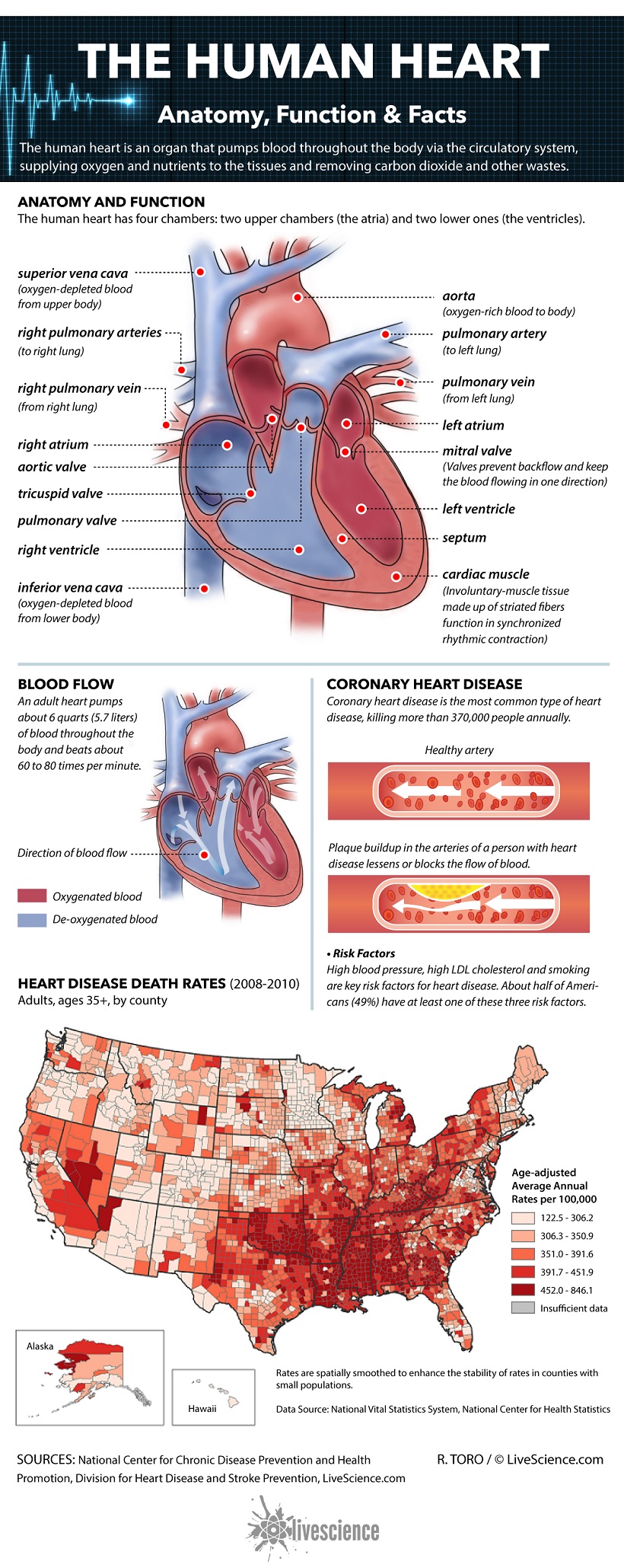
The heart is a muscular organ about the size of a closed fist that functions as the bodys circulatory pump. It takes in deoxygenated blood through the veins and delivers it to the lungs for oxygenation before pumping it into the various arteries (which provide oxygen and nutrients to body tissues by transporting the blood throughout the body).
The heart is located in the thoracic cavity medial to the lungs and posterior to the sternum.
On its superior end, the base of the heart is attached to the aorta, pulmonary arteries and veins, and the vena cava.
The inferior tip of the heart, known as the apex, rests just superior to the diaphragm.
The base of the heart is located along the bodys midline with the apex pointing toward the left side. Because the heart points to the left, about 2/3 of the hearts mass is found on the left side of the body and the other 1/3 is on the right.
Anatomy of the Heart Pericardium
The heart sits within a fluid-filled cavity called the pericardial cavity. The walls and lining of the pericardial cavity are a special membrane known as the pericardium. Pericardium is a type of serous membrane that produces serous fluid to lubricate the heart and prevent friction between the ever beating heart and its surrounding organs.
Besides lubrication, the pericardium serves to hold the heart in position and maintain a hollow space for the heart to expand into when it is full.
The pericardium has 2 layersa visceral layer that covers the outside of the heart and a parietal layer that forms a sac around the outside of the pericardial cavity.
Structure of the Heart Wall
The heart wall is made of 3 layers: epicardium, myocardium and endocardium.
Epicardium. The epicardium is the outermost layer of the heart wall and is just another name for the visceral layer of the pericardium. Thus, the epicardium is a thin layer of serous membrane that helps to lubricate and protect the outside of the heart. Below the epicardium is the second, thicker layer of the heart wall: the myocardium.
Myocardium. The myocardium is the muscular middle layer of the heart wall that contains the cardiac muscle tissue. Myocardium makes up the majority of the thickness and mass of the heart wall and is the part of the heart responsible for pumping blood. Below the myocardium is the thin endocardium layer.
Endocardium. Endocardium is the simple squamous endothelium layer that lines the inside of the heart. The endocardium is very smooth and is responsible for keeping blood from sticking to the inside of the heart and forming potentially deadly blood clots.
The thickness of the heart wall varies in different parts of the heart. The atria of the heart have a very thin myocardium because they do not need to pump blood very faronly to the nearby ventricles.
The ventricles, on the other hand, have a very thick myocardium to pump blood to the lungs or throughout the entire body.
The right side of the heart has less myocardium in its walls than the left side because the left side has to pump blood through the entire body while the right side only has to pump to the lungs.
Chambers of the Heart
The heart contains 4 chambers: the right atrium, left atrium, right ventricle, and left ventricle. The atria are smaller than the ventricles and have thinner, less muscular walls than the ventricles.
The atria act as receiving chambers for blood, so they are connected to the veins that carry blood to the heart. The ventricles are the larger, stronger pumping chambers that send blood out of the heart.
The ventricles are connected to the arteries that carry blood away from the heart.
The chambers on the right side of the heart are smaller and have less myocardium in their heart wall when compared to the left side of the heart.
This difference in size between the sides of the heart is related to their functions and the size of the 2 circulatory loops.
The right side of the heart maintains pulmonary circulation to the nearby lungs while the left side of the heart pumps blood all the way to the extremities of the body in the systemic circulatory loop.
Valves of the Heart
The heart functions by pumping blood both to the lungs and to the systems of the body. To prevent blood from flowing backwards or regurgitating back into the heart, a system of one-way valves are present in the heart.
The heart valves can be broken down into two types: atrioventricular and semilunar valves.
Atrioventricular valves. The atrioventricular (AV) valves are located in the middle of the heart between the atria and ventricles and only allow blood to flow from the atria into the ventricles. The AV valve on the right side of the heart is called the tricuspid valve because it is made of three cusps (flaps) that separate to allow blood to pass through and connect to block regurgitation of blood.
The AV valve on the left side of the heart is called the mitral valve or the bicuspid valve because it has two cusps. The AV valves are attached on the ventricular side to tough strings called chordae tendineae.
The chordae tendineae pull on the AV valves to keep them from folding backwards and allowing blood to regurgitate past them. During the contraction of the ventricles, the AV valves look like domed parachutes with the chordae tendineae acting as the ropes holding the parachutes taut.
Semilunar valves. The semilunar valves, so named for the crescent moon shape of their cusps, are located between the ventricles and the arteries that carry blood away from the heart. The semilunar valve on the right side of the heart is the pulmonary valve, so named because it prevents the backflow of blood from the pulmonary trunk into the right ventricle. The semilunar valve on the left side of the heart is the aortic valve, named for the fact that it prevents the aorta from regurgitating blood back into the left ventricle. The semilunar valves are smaller than the AV valves and do not have chordae tendineae to hold them in place. Instead, the cusps of the semilunar valves are cup shaped to catch regurgitating blood and use the bloods pressure to snap shut.
Conduction System of the Heart
The heart is able to both set its own rhythm and to conduct the signals necessary to maintain and coordinate this rhythm throughout its structures.
About 1% of the cardiac muscle cells in the heart are responsible for forming the conduction system that sets the pace for the rest of the cardiac muscle cells.
The conduction system starts with the pacemaker of the hearta small bundle of cells known as the sinoatrial (SA) node.
The SA node is located in the wall of the right atrium inferior to the superior vena cava.
The SA node is responsible for setting the pace of the heart as a whole and directly signals the atria to contract.
The signal from the SA node is picked up by another mass of conductive tissue known as the atrioventricular (AV) node.
The AV node is located in the right atrium in the inferior portion of the interatrial septum.
The AV node picks up the signal sent by the SA node and transmits it through the atrioventricular (AV) bundle.
The AV bundle is a strand of conductive tissue that runs through the interatrial septum and into the interventricular septum. The AV bundle splits into left and right branches in the interventricular septum and continues running through the septum until they reach the apex of the heart.
Branching off from the left and right bundle branches are many Purkinje fibers that carry the signal to the walls of the ventricles, stimulating the cardiac muscle cells to contract in a coordinated manner to efficiently pump blood out of the heart.
Physiology of the Heart Coronary Systole and Diastole
At any given time the chambers of the heart may be found in one of two states:
Systole. During systole, cardiac muscle tissue is contracting to push blood out of the chamber.
Diastole. During diastole, the cardiac muscle cells relax to allow the chamber to fill with blood. Blood pressure increases in the major arteries during ventricular systole and decreases during ventricular diastole.
This leads to the 2 numbers associated with blood pressuresystolic blood pressure is the higher number and diastolic blood pressure is the lower number.
For example, a blood pressure of 120/80 describes the systolic pressure (120) and the diastolic pressure (80).
The Cardiac Cycle
The cardiac cycle includes all of the events that take place during one heartbeat.
There are 3 phases to the cardiac cycle: atrial systole, ventricular systole, and relaxation.
Atrial systole: During the atrial systole phase of the cardiac cycle, the atria contract and push blood into the ventricles. To facilitate this filling, the AV valves stay open and the semilunar valves stay closed to keep arterial blood from re-entering the heart.
The atria are much smaller than the ventricles, so they only fill about 25% of the ventricles during this phase. The ventricles remain in diastole during this phase.
Ventricular systole: During ventricular systole, the ventricles contract to push blood into the aorta and pulmonary trunk. The pressure of the ventricles forces the semilunar valves to open and the AV valves to close.
This arrangement of valves allows for blood flow from the ventricles into the arteries. The cardiac muscles of the atria repolarize and enter the state of diastole during this phase.
Relaxation phase: During the relaxation phase, all 4 chambers of the heart are in diastole as blood pours into the heart from the veins. The ventricles fill to about 75% capacity during this phase and will be completely filled only after the atria enter systole.
The cardiac muscle cells of the ventricles repolarize during this phase to prepare for the next round of depolarization and contraction. During this phase, the AV valves open to allow blood to flow freely into the ventricles while the semilunar valves close to prevent the regurgitation of blood from the great arteries into the ventricles.
Blood Flow through the Heart
Deoxygenated blood returning from the body first enters the heart from the superior and inferior vena cava. The blood enters the right atrium and is pumped through the tricuspid valve into the right ventricle.
From the right ventricle, the blood is pumped through the pulmonary semilunar valve into the pulmonary trunk.
The pulmonary trunk carries blood to the lungs where it releases carbon dioxide and absorbs oxygen. The blood in the lungs returns to the heart through the pulmonary veins.
From the pulmonary veins, blood enters the heart again in the left atrium.
The left atrium contracts to pump blood through the bicuspid (mitral) valve into the left ventricle. The left ventricle pumps blood through the aortic semilunar valve into the aorta.
From the aorta, blood enters into systemic circulation throughout the body tissues until it returns to the heart via the vena cava and the cycle repeats.
The Electrocardiogram
The electrocardiogram (also known as an EKG or ECG) is a non-invasive device that measures and monitors the electrical activity of the heart through the skin. The EKG produces a distinctive waveform in response to the electrical changes taking place within the heart.
The first part of the wave, called the P wave, is a small increase in voltage of about 0.1 mV that corresponds to the depolarization of the atria during atrial systole.
The next part of the EKG wave is the QRS complex which features a small drop in voltage (Q) a large voltage peak (R) and another small drop in voltage (S).
The QRS complex corresponds to the depolarization of the ventricles during ventricular systole. The atria also repolarize during the QRS complex, but have almost no effect on the EKG because they are so much smaller than the ventricles.
The final part of the EKG wave is the T wave, a small peak that follows the QRS complex. The T wave represents the ventricular repolarization during the relaxation phase of the cardiac cycle.
Variations in the waveform and distance between the waves of the EKG can be used clinically to diagnose the effects of heart attacks, congenital heart problems, and electrolyte imbalances.
Heart Sounds
The sounds of a normal heartbeat are known as lubb and dupp and are caused by blood pushing on the valves of the heart. The lubb sound comes first in the heartbeat and is the longer of the two heart sounds.
The lubb sound is produced by the closing of the AV valves at the beginning of ventricular systole. The shorter, sharper dupp sound is similarly caused by the closing of the semilunar valves at the end of ventricular systole.
During a normal heartbeat, these sounds repeat in a regular pattern of lubb-dupp-pause. Any additional sounds such as liquid rushing or gurgling indicate a structure problem in the heart.
The most likely causes of these extraneous sounds are defects in the atrial or ventricular septum or leakage in the valves.
Cardiac Output
Cardiac output (CO) is the volume of blood being pumped by the heart in one minute.
The equation used to find cardiac output is: CO = Stroke Volume x Heart Rate
Stroke volume is the amount of blood pumped into the aorta during each ventricular systole, usually measured in milliliters.
Heart rate is the number of heartbeats per minute. The average heart can push around 5 to 5.5 liters per minute at rest.
Here are links to some popular and reputable Websites, Foundations, Organizations, Associations, Societies, Etc about the heart and where you can find additional information.
HeartSite.com
This site has to rate in the top as one of the best learning sites I have seen. If you want to learn and better understand the heart and all about it, browse this site, you will be amazed.
This web site was designed to provide information to patients who are being evaluated and treated for a heart-related complaint. All contents are reviewed by physicians to ensure accuracy. Our objective is to educate. The information on this web site is meant to supplement and NOT to replace those obtained from your personal physician. Please consult your physician because a specific disease, test or treatment may not be applicable to your case. To facilitate understanding, tools and lectures were designed from the ground up instead of scanning in traditional textbooks or videotaping a slide presentation and then modifying it for use on the Internet. This site includes panoramic views, 3D animation, online lectures, narrations, streaming instructional video, and easy to understand animated examples.
All of the material on this web site is made up of original work designed by the web site team and reviewed by the Medical Editor.
Heart Disease Articles
Heart disease is the number one killer of adults in the United States. In fact, one in every three deaths is from heart disease and stroke, equal to 2,200 deaths per day.
Taking care of your heart is essential for a long life. You can treat your ticker right by eating healthy, staying active and managing your stress.
Here are 80+ articles relating to about every aspect of Heart Disease you can imagine with a multitude of information.
Also some Heart Disease Faqs and more. Source:Sharecare.com (All Your Health In One Place).
The Children's Heart Foundation
The Children's Heart Foundation is a not-for-profit organization that funds the most promising research to advance the diagnosis, treatment and prevention of congenital heart defects.
The Heart Foundation
The Heart Foundations mission is to save lives by educating the public about heart disease, promoting early detection, and supporting the research of world-renowned cardiologist Dr. P.K. Shah in the quest to find more effective ways to prevent and treat the #1 cause of death in the U.S. We dedicate ourselves to this mission in memory of Steven S. Cohen, an incredible husband, father, son, brother and friend.
On December 7, 1995, Steven Cohen came home from work, changed his clothes, kissed his wife and daughters (ages 2 and 5) and walked out the door to play his weekly basketball game with friends. Neither Steve nor his wife Eva could know what would follow. During the basketball game this healthy-looking, 35-year-old man complained of not feeling well. He left the court, sat down and suffered a massive heart attack that took his life.
American Heart Association
The American Heart Association is the nations oldest and largest voluntary organization dedicated to fighting heart disease and stroke. Founded by six cardiologists in 1924, our organization now includes more than 22.5 million volunteers and supporters. We fund innovative research, fight for stronger public health policies, and provide critical tools and information to save and improve lives.
Our nationwide organization includes 156 local offices and more than 3,000 employees. We moved our national headquarters from New York to Dallas in 1975 to be more centrally located. The American Stroke Association was created as a division in 1997 to bring together the organizations stroke-related activities.
To read more about the AHA Go Here.
World Heart Federation
Striving for a 25% reduction by 2025 in premature deaths from cardiovascular disease around the world.
The World Heart Federation is dedicated to leading the global fight against cardiovascular disease (CVD), including heart disease and stroke. With a focus on low- and middle-income countries, we are the only global advocacy and leadership organization bringing together the CVD community to help people lead heart-healthy lives.
International Children's Heart Foundation
The mission of the International Childrens Heart Foundation (ICHF) is to bring the skills, technology and knowledge to cure and care for children with congenital heart disease in developing nations. ICHF does this regardless of country of origin, race, religion or gender. Our goal is to make the need for ICHF obsolete. We work toward this goal through our medical mission trips, where we operate on children and educate local healthcare professionals.
Heart Failure Society of America
The Heart Failure Society of America, Inc. (HFSA) represents the first organized effort by heart failure experts from the Americas to provide a forum for all those interested in heart function, heart failure, and congestive heart failure (CHF) research and patient care.
The mission of HFSA is to:
- Promote research related to all aspects of heart failure and to provide a forum for presentation of basic, clinical, and population-based research.
- Educating health care professionals through programs, publications, and other media in the areas of basic science, clinical medicine, patient management, and social, ethical and economic issues to enable them to diagnose and treat heart failure and concomitant medical conditions more effectively.
- Encourage primary and secondary preventive measures to reduce the incidence of heart failure; to serve as a resource for government, private industry, and health care providers to facilitate the establishment of programs and policies that will better serve the patient.
- Enhance quality and duration of life in those with heart failure.
- Promote and facilitate the formal training of physicians, scientists, and allied health care providers in the field of heart failure.
InterAmerican Heart Foundation
Heart attack and blood vessel diseases, including high blood pressure and stroke, know no boundaries.
In almost all countries of the Americas, these diseases are the number one cause of death. The highest mortality rates for all cardiovascular diseases are found in the English-speaking Caribbean, the USA, Canada, Argentina, Chile and Uruguay. Death rates from cardiovascular diseases are on the rise in Central American and Latin Caribbean regions.
More people are using tobacco. Diets have more fat. And physical work is being replaced by labor-saving devices leading to sedentary lifestyles and weight gain. The combination of bad lifestyle habits and urbanization, over time, leads to high cholesterol, diabetes, high blood pressure, and the premature development of atherosclerosis.
The result can be heart attack, heart failure, stroke, peripheral vascular disease, and other vascular problems. Medical treatment requires huge expenditures for medical and hospital care, depleting economic resources at all levels of the countries of the Americas.
International Society for Heart Research
Our mission is to promote the discovery and dissemination of knowledge in the cardiovascular sciences on a world-wide basis through publications, congresses and other media. Our goals are:
- To promote the exchange of ideas on a world-wide basis between scientists and clinicians interested in all aspects of cardiovascular biology and medicine.
- To promote discovery and dissemination of knowledge in all areas of cardiovascular biology and medicine.
- To organize and support national and international congresses devoted to all aspects of cardiovascular research.
- To provide an international forum for discussion of problems and controversies at the cutting edge of cardiovascular research.
- To publish a world class journal devoted to advances in cardiovascular research.
Women Heart
The National Coalition for Women with Heart Disease.
WomenHeart's mission is to improve the health and quality of life of women living with or at risk of heart disease, and to advocate for their benefit.
CongenitalHeartDefects.com
Congenital heart defects are responsible for more deaths in the first year of life than any other birth defects, the National Institutes of Health reports. This site is devoted to providing information to members of the world-wide congenital heart defect (CHD) community.
We do not provide medical advice, but we do have information which can supplement what doctors and other professionals provide.
Whether you are looking for definitions of medical terms, hospitals caring for people with CHDs, other people with whom you can network and share experiences, links to support groups, articles regarding CHDs, or resources for the CHD community, you have come to the right place!
Heart Foundation
The Heart Foundation is a charity dedicated to fighting the single biggest killer of Australians heart disease. For over 50 years, weve led the battle to save lives and improve the heart health of all Australians. Our sights are set on a world where people dont suffer or die prematurely because of heart disease.
Go Red for Women
Do you know what causes heart disease in women? What about the survival rate? Or whether women of all ethnicities share the same risk?
The fact is: Heart disease is the No. 1 killer of women, causing 1 in 3 deaths each year. Thats approximately one woman every minute!
But it doesnt affect all women alike, and the warning signs for women arent the same in men. Whats more: These facts only begin to scratch the surface.
There are several misconceptions about heart disease in women, and they could be putting you at risk. The American Heart Associations Go Red For Women movement advocates for more research and swifter action for womens heart health for this very reason. In this section, well arm you with the facts and dispel some myths because the truth can no longer be ignored.
Million Hearts
Heart disease and stroke are the first and fifth leading causes of death in the United States. Every 43 seconds, someone in the United States has a heart attack, many of them fatal. On average, one American dies from stroke every 4 minutes.
Million Hearts is a national initiative with an ambitious goal to prevent 1 million heart attacks and strokes by 2017. The Centers for Disease Control and Prevention and the Centers for Medicare & Medicaid Services co-lead the initiative on behalf of the U.S. Department of Health and Human Services.
Adult Congenital Heart Association
Supporting Patients and Families
Congenital heart disease is survivable, manageable, yet in the routine years between infancy and adulthood, sometimes forgettable. The Adult Congenital Heart Association is a resource, advocate and knowledge base for everyone affected.
American College of Cardiology
The ACC's Strategic Plan charts the course for the College over the next five years. The plan leverages the College's role as the professional home for the cardiovascular care team and positions the College and its members for success in meeting the Triple Aim of improving cardiovascular health through lower costs, better health and better outcomes.
Children's Heart Association
When a child is diagnosed with a heart problem it is a difficult time for the whole family. The Childrens Heart Association would like to help.
What do we do?
- Support heart children and young people, their families and friends.
- Raise funds to support our activities & to buy equipment.
- Make life a little easier during hospital visits & stays.
- Share information and tips.
- Have fun.
Kids With Heart
Kids With Heart National Association for Children's Heart Disorders was founded in 1985 as a local support group for families affected by congenital heart defects in Northern Wisconsin. In 1991, Kids With Heart went National and the name was officially changed to Kids With Heart National Association for Children's Heart Disorders, Inc. It was the first National organization dedicated to providing support, education, and awareness for congenital heart defects. Kids With Heart NACHD, Inc is a non-profit organization and has been run by volunteers since the very beginning. Therefore, we have no salaries to pay, no rent to pay, no senseless overhead costs.
Little Hearts, Inc.
Little Hearts, Inc. is a national organization providing support, education, resources, networking, and hope to families affected by congenital heart defects. Membership consists of families nationwide who have or are expecting a child with a congenital heart defect.
Our mission is to offer support, education, and hope to families affected by congenital heart defects through our support services, and to promote public awareness for this #1 birth defect. Read More.
National Heart, Lung, and Blood Institute The National Heart, Lung, and Blood Institute (NHLBI) provides global leadership for a research, training, and education program to promote the prevention and treatment of heart, lung, and blood diseases and enhance the health of all individuals so that they can live longer and more fulfilling lives.
The NHLBI stimulates basic discoveries about the causes of disease, enables the translation of basic discoveries into clinical practice, fosters training and mentoring of emerging scientists and physicians, and communicates research advances to the public. It creates and supports a robust, collaborative research infrastructure in partnership with private and public organizations, including academic institutions, industry, and other government agencies.
The Institute collaborates with patients, families, health care professionals, scientists, professional societies, patient advocacy groups, community organizations, and the media to promote the application of research results and leverage resources to address public health needs. The NHLBI also collaborates with international organizations to help reduce the burden of heart, lung, and blood diseases worldwide.
Pediatric Congenital Heart Association
The Pediatric Congenital Heart Associations mission is to Conquer Congenital Heart Disease. We are accomplishing this through collaboration with patients, parents, providers, and partner organizations in order to effectively advocate for improved quality and outcomes through CHD education, research and awareness.
Founded in 2013, the Pediatric Congenital Heart Association quickly filled a niche as the voice of the congenital heart patient. Our Leadership includes more than 30 patients, parents and providers from across the country making up our Board, volunteer committees and our Medical Advisory Board.
We are founded on the key purpose to be the resounding voice of the pediatric patient population and are accomplishing this through collaboration with patients, parents, providers, and partner organizations. We are improving quality and outcomes through CHD education, research and awareness, all while reducing the impact of congenital heart disease while striving to realize a world free from it.
Mend a Heart Foundation
In support of all children born with congenital heart defects and medical practitioners and researchers who advance congenital heart defect research, the Mend a Heart Foundation supports initiatives that extend and enrich the lives of heart kids.
Heart Rhythm Society
In pursuit of its vision to end death and suffering due to heart rhythm disorders, HRS overarching goals will be to secure its financial future through focus on increasing reach, engaging members, and building its brand as the recognized authority for heart rhythm care.
World Heart Failure Society
The WHFS is a non-profit organization and a professional society. We would like to bring together medical, allied health and research professionals from all parts of the world who are involved in the field of heart failure. As heart failure is a complex syndrome that encompasses many facets of management, it typically involves all subspecialties of cardiovascular medicine. With the rapid evolution in the management of heart failure, we hope that the WHFS will provide a platform of knowledge transfer, exchange of ideas and experiences, professional education and foster collaborative search on a global basis.
Heart Failure Online
Heart Failure Online is dedicated to providing information regarding heart failure to patients and their families.
We have worked hard to break down the components of heart failure, starting with an overview of the organ systems involved and progressing through how each organ system is affected, methods of prevention, treatment options, and much more.
Caregivers of people with heart failure are usually spouses, children, grandchildren, siblings, parents, or family friends.
Sometimes a carer may be provided by the national health system or employed privately.
Being a caregiver for someone with a chronic medical condition such as heart failure can be both physically exhausting and emotionally draining at times.
You may find yourself pulled in many directions as you provide practical help to your partner or relative, as well as experiencing a range of mixed emotions yourself.
Becoming a caregiver requires a long-term commitment of time and energy, but with a clear plan and good support it can also be a fulfilling and rewarding experience.
In this section, we look at the role of a caregiver and the special challenges that you face in caring for your partner or relative, whilst providing you with some ideas and helpful tips to make your role easier. You can adapt any suggestions to your own circumstances and preferences, as your needs and experiences are unique to you.
Understanding Your Role
Being a caregiver may mean that you have to play many roles all at once, including being a friend, confidant, nurse and assistant. Your role as caregiver may happen gradually, over a period of time, or it can happen suddenly, if your partner or relative has a heart attack for example.
Your role will involve helping, supporting and coaching your partner or relative to help adjust their lifestyle to fit with their new life managing their heart failure.
You are likely to have the largest impact on their success in working with their healthcare team and following their instructions.
It is essential that you are involved because for many people managing their heart failure, the caregiver is the most important person involved in their care.
You should aim to help your partner or relative to learn and accept that the self-management side of living with heart failure is down to them whilst trying not to be over protective.
Being a caregiver is about being the patients coach and not feeling obliged to do things. It is about enabling the patient to self-manage.
Your role as carer may not be permanent and the help and support your partner or relative needs at the moment may get less with time.
Click on the links below to find out more about the help and support you can give your partner or relative.
Simple Things You Can Do To Help
Managing heart failure can be a challenge, and your partner or relative may not be able to do it alone. Physically, they may not be able to handle all the tasks they once could.
The diet and lifestyle changes their doctor recommends may be confusing.
On top of this, the number of medicines they may need to take can be overwhelming at first.
You and other family members and friends can provide a much-needed source of care and support in these areas.
Click below to read about some simple things that you can do to help your partner or relative adjust to life with heart failure.
Understanding Their Emotions
It is common for someone to feel depressed or anxious after learning that they have a chronic disease such as heart failure.
For most people, these feelings get better and fade as they learn that the condition can be managed.
For others, however, these negative feelings may continue or even get worse over time. It is important to take some positive steps to help your partner or relative manage these feelings.
Click on the links below to find out more about recognizing depression and anxiety in your partner or relative and suggestions of how you could help.
How You May Be Feeling
As a caregiver, it’s all too easy to put all your energy, efforts and attention into caring for your partner or relative at the expense of your own needs and feelings. As everyone is unique, emotional responses to situations will very much depend on the individual, but you may have experienced some of the feelings below in your role as caregiver:
You may feel that you always have to appear positive, cheerful and optimistic, even when you are struggling with certain issues.
You may tend to bottle feelings up which may eventually lead to you experiencing ill health or high levels of depression.
You may feel anxious because you are not sure what is happening to your partner or relative, or because you feel responsible for their health. If your partner or relative is also very anxious, it’s hard for you not to share their anxiety too.
Circumstances could make you feel resentful. You may feel frustrated about the changes in your life or resentful at your partner or relative for being “the cause” of these changes. Also, if your partner or relative is being irritable or uncooperative, you may feel resentful and unappreciated for all that you are doing.
You may feel angry because your partner or relative has heart failure. You may wonder why it happened to them and feel angry at life, or you could feel angry with them for not preventing it by stopping smoking or failing to follow medical advice properly. You may feel guilty for having these feelings of anger and resentment.
You may think that you have to be the strong one and that having any of the above feelings means you are betraying your partner or relative in some way.
There may be occasions when you experience a range of emotions in a short period of time. One day you may feel positive and in control, and the next day quite depressed and uncertain. At times, you may feel very isolated and feel that nobody else can possibly understand what you are going through.
Please be reassured, you are not the only one going through these emotions. You may find it helpful to talk to people in similar situations. Click Here for more information on support networks for caregivers.
Support Networks
In the same way that you are providing invaluable help and support to your partner or relative to manage their heart failure, you need just as much support in your role as a caregiver. You may feel very alone at times as you go about coping with everything, but there are many support systems available to you:
TALK to someone. Talking may be the most simple and effective support system that you need. If you don't feel you can talk to your partner or relative about certain things then talk to a family member, a trusted friend, a doctor or nurse, or a trained counsellor.
Seek help from family and friends, a volunteer group or help provided from your local health authority. You cannot and should not have to do everything on your own. Speak to your own doctor, or your partner or relative's doctor, to see what extra help may be available to you through services in your area.
Join a caregiver support group. Just as your partner or relative can use a patient support group, you can consider joining a caregiver support group to spend time with others in similar situations. You can find out about appropriate support groups by speaking to your doctor or nurse, asking at your local library or searching online.
Here are a few more suggestions that could help you feel more in control:
Learn more about heart failure. The more you know, the more in control you are likely to feel.
Make time for yourself and the activities you enjoy.
Plan for the future. If you are feeling uncertain about the future, you may get some peace of mind from working with your partner or relative to make sure that finances, wills and insurance policies are in order.
This may also provide an opportunity to discuss future care plans with your partner or relative.
Financial Concerns
It is possible that a diagnosis of heart failure may raise important financial concerns for you and your partner or relative.
Healthcare systems differ between countries and everyone's situation is different. However, the following suggestions may help you start dealing with any financial matters you need to consider:
Understand your partner or relative's health insurance plan(s). If your partner or relative has a health insurance plan, clarify with them what is covered and what effect their heart failure diagnosis may have on the policy. It’s important to bear in mind that some of the newest advances in treatment for heart failure (new medicines, procedures) are still technically “under investigation” in clinical studies, so an insurance plan may not cover them. Speak with your partner or relative's doctor about any questions you may have in this area or go directly to the healthcare insurer.
If necessary, seek financial assistance for treatment costs. It’s essential for heart failure patients to take all their medicines as prescribed. You can speak to your doctor or nurse about this or they may be able to refer you to a social services department that can help with or advise on any financial issues.
Look at household finances. If the person with heart failure is the main breadwinner or contributes significantly to the household income, then you and your family may well have to make some financial adjustments if they are not able to work to the extent they were before. If your partner or relative has disability insurance you may be able to make a claim on this.
Planning For The Future
You may be very reluctant to speak to your partner or relative about what to expect in the future with their health.
These issues are extremely difficult to raise, especially when you are trying to be positive.
However, it is very important that you and your partner or relative bear in mind that planning for the future is not the same as giving up.
This is actually a way for you both to cope and control the situation better and make things easier for all concerned.
Although it's difficult to initiate, dealing with these issues can bring a great deal of peace of mind to you and your family, leaving you to focus your energies on living a full life.
As soon as you feel up to it, talk with your partner or relative about drawing up an Advance Care Directive.
This involves looking at preparing documents, which explain your partner or relative's wishes for medical care in the future, in the event he or she becomes unable to communicate. Click Here for more information on planning for your partner or relatives end of life.
You may find this is also a good time to make sure that financial matters, including wills and life insurance policies, are in order.
Message from Site Owner and Webmaster
All health and health related information contained within the Your Heart and Health Medical Information Website is intended to be general in nature and should not be used as a substitute for a visit with a health care professional. The advice is intended to offer only a general basis for individuals to discuss their medical condition with their health care provider. Your health care provider should be consulted regarding matters concerning the medical condition, treatment and needs of you and your family.
For medical matters, please contact your doctors office by telephone, in emergency situations, call 911.
Please read the rest of the Disclaimers and Private Policy Go Here.
If you are seeing this, you have attempted to link to the UpToDate widget but are experiencing a problem. Please visit UpToDate for more information.
Access the FREE Beyond the Basics (English) patient content in UpToDate by simply typing a term or phrase in the search box. Look up your specific question anything from a symptom you're concerned about to a condition or treatment you've been discussing with your healthcare provider.
Most of our widgets are built using Adobe Flash.
You will need the FREE Adobe Flash Player plug-in to view them.
Type a medical word, phrase, question, etc and click GO
CDC is the nation's health protection agency, working 24/7 to protect America from health and safety threats, both foreign and domestic. CDC increases the health security of our nation.
Founded by Lynn B Goddess in April 2000 for Stroke Research in Women as a legacy to her mother who died of complications from stroke. Lynn's journey meeting the demands and challenges of her mother's illness dramatizes the overwhelming gap that exist between perception and reality when it comes to stroke in women.
Read the article "What Women need to know about stroke" in StrokeSmart Magazine Click Here
Get a brief definition of dozens of cardiovascular terms from our A-Z Heart and Stroke Encyclopedia and get links to in-depth information.
The mission of the Division for Heart Disease and Stroke Prevention (DHDSP) is to provide public health leadership to improve cardiovascular health for all, reduce the burden, and eliminate disparities associated with heart disease and stroke.
Atrial fibrillation (AFib) not caused by a heart valve problem is a type of irregular heartbeat that was estimated to affect approximately 6.4 million people in the U.S. in 2014.
For some quick facts about Afib and Stroke Click Here
An up to date web site, you can find the health information you are looking for here.
Take the approximately 2.5 minute tour and learn more. Start Here
It's not just a man's disease.
1 in 3 women die of heart disease and stroke.
But it can be prevented.
Join us to receive simple, inspiring tips to live a heart-healthy life. Learn More
AHA Compliant Online CPR Certification that over 600,000 professionals trust. Means you can train and test online and print your CPR certificate instantly! Get Yours
Improve your life and the lives of others when you join the AHA/ASA Support Network
Share your experience, give and get emotional support.
Get quick answers to quick questions. Read More
The COPD Foundation is a non-profit organization created in 2004,
Established to undertake initiatives that result in expanded services for COPD and improve the lives of individuals affected by COPD. Learn More
One out of every 10 cancer patients who receives chemotherapy gets an infection that requires a hospital visit. More Info
Start here to learn the basics of HIV and AIDS and learn how to take steps to prevent transmission and protect yourself from HIV.
50% of young people who have HIV don't know they have it. Get the facts HERE
About 1 of 3 adults, 67 million people have high blood pressure.
High blood pressure makes your heart work too hard and increases your risk of heart disease and stroke. Learn More
Very interesting and detailed charts and graphs for states, federal regions, and territories. More Info
True stories from previous smokers who have quit after having serious heart problems. A lot of tips and suggestions to help you quit. Click Here
People with ADHD may have trouble paying attention, controlling impulsive behaviors, or be overly active
ADHD can't be cured, but it can be successfully managed and some symptoms may improve as the child ages. Read More
Watch health videos on topics such as anatomy and body systems, surgical procedures and health news.
Test your knowledge with interactive tutorials and games.
Check your health with calculators and quizzes. Try It